临床荟萃 ›› 2025, Vol. 40 ›› Issue (6): 532-536.doi: 10.3969/j.issn.1004-583X.2025.06.009

• 论著 • 上一篇    下一篇

以癫痫发作为首发临床表现的急性肺栓塞1例并文献复习

王肖肖1(), 胡彦峰2, 祁秀峰1, 赵俊杰1   

  1. 1.邯郸市第一医院 神经内科,河北 邯郸 056000
    2.邯郸市中心医院 呼吸内科,河北 邯郸 057150
  • 收稿日期:2025-01-03 出版日期:2025-06-20 发布日期:2025-07-01
  • 通讯作者: 王肖肖 E-mail:779143771@qq.com
  • 基金资助:
    邯郸市科学技术研究与发展计划项目——丛集性癫痫发作的临床分析及预后(23422083279)

Acute pulmonary embolism with epilepsy attack as the first clinical manifestation: A case report and literature review

Wang Xiaoxiao1(), Hu Yanfeng2, Qi Xiufeng1, Zhao Junjie1   

  1. 1. Department of Neurology, Handan First Hospital, Handan 056000, China
    2. Department of Respiratory Medicine, Handan Central Hospital, Handan 057150, China
  • Received:2025-01-03 Online:2025-06-20 Published:2025-07-01
  • Contact: Wang Xiaoxiao E-mail:779143771@qq.com

摘要:

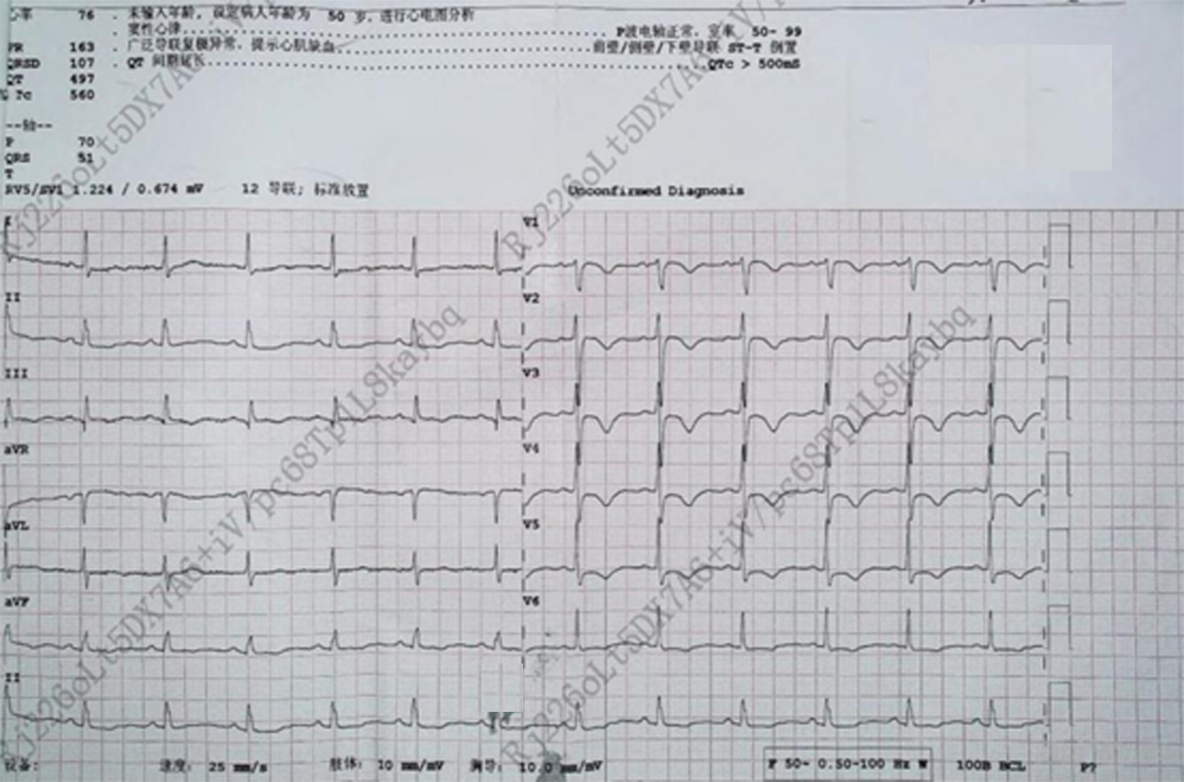
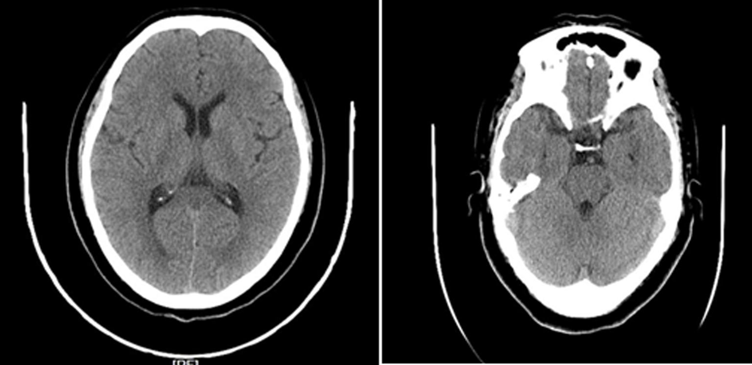
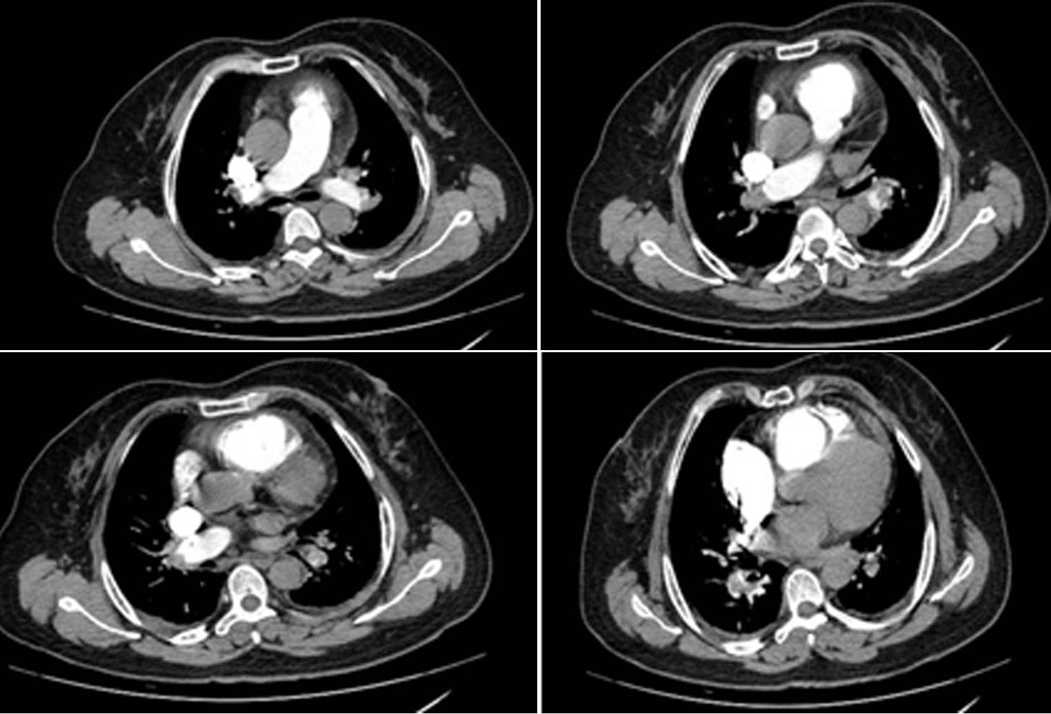
目的 探讨以癫痫发作为首发临床表现的急性肺栓塞的临床特征及诊疗思路。方法 回顾性分析1例以癫痫发作为首发临床表现的急性肺栓塞患者的诊治过程, 并复习相关文献。结果 患者为59岁女性,癫痫发作表现为意识丧失,摔倒在地,双眼上吊,牙关紧闭,伴舌咬伤,小便失禁,无四肢抽搐,持续约10 min后意识转清,醒后自觉头晕,双下肢无力,不能回忆,非夜间发作;D-二聚体高,血气分析示低氧血症、呼吸性碱中毒;心电图示窦性心律,SIQT,广泛T波低平、倒置;心脏彩色超声示肺动脉中度高压,左室舒张功能减低、收缩功能正常;双下肢血管彩色超声示右侧肌间静脉血栓形成,左侧肌间静脉增宽;肺CTA示双侧多发肺动脉栓塞;肺动脉主干轻度加宽。结论 急性肺栓塞症状呈多样性,隐匿性,当遇到既往无脑损伤及癫痫发作病史,首次癫痫发作,D-二聚体高、心电图提示:窦性心律,SIQT,广泛T波低平、倒置,警惕急性肺栓塞的诊断,以避免误诊,并及时治疗,改善预后。

关键词: 癫痫, 肺栓塞, 心电描记术, 肺CTA

Abstract:

Objective To explore the clinical characteristics and diagnosis and treatment of acute pulmonary embolism (APE) with epilepsy attack as the first clinical manifestation. Methods The diagnosis and treatment of a case of APE with epilepsy attack as the first clinical manifestation were retrospectively analyzed, and the related literatures were reviewed. Results A 59-year-old female patient exhibited non-night epilepsy attack, manifesting as loss of consciousness, falling to the ground, hanging eyes, tight teeth, tongue biting, and incontinence, without limb twitching. She returned to clear consciousness after 10 minutes. After waking up, she felt dizzy, weak in both lower limbs, and unable to recall. Laboratory testing suggested elevated D-dimer, and blood gas analysis showed hypoxemia and respiratory alkalosis. Electrocardiogram showed sinus rhythm, SIQT, and extensive low, flat, and inverted T waves. Cardiac color Doppler showed moderate pulmonary hypertension, reduced left ventricular diastolic function, and normal contraction function. Color Doppler of lower limbs showed right intermuscular venous thrombosis, and widened left intermuscular veins. The computed tomography pulmonary angiogram (CTPA) showed multiple pulmonary embolisms on both sides, and a mild widening of the main trunk of pulmonary artery. Conclusion The symptoms of APE are diverse and occult. An alert of APE is made in case of a history of epilepsy attack without brain injuries, elevated D-dimer, and sinus rhythm SIQT, and extensive low, flat, and inverted T waves on electrocardiogram. Cautions should be made to prevent misdiagnoses. A timely treatment favors the prognosis.

Key words: epilepsy, pulmonary embolism, electrocardiography, lung CTA

中图分类号: